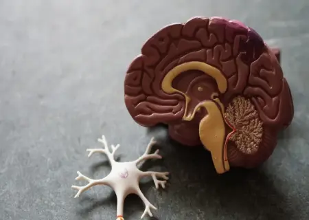
The brain is a complex organ that acts as the control center of the body. As a component of the central nervous system, the brain sends, receives, processes, and directs sensory information. The brain is split into left and right hemispheres by a band of fibres called the corpus callosum.
DIVISION OF BRAIN:
There are three major divisions of the brain, with each division performing specific functions. The major divisions of the brain are
1) Forebrain ( prosencephalon)
2) Midbrain (mesencephalon)
3) Hindbrain (rhombencephalon)
FOREBRAIN (PROSENCEPHALON):
The forebrain plays a central role in the processing of information related to cognitive activities, sensory and associative functions, and voluntary motor activities.
It includes
1) Telencephalon
2) Diencephalon
Telencephalon contains cerebral hemisphere. The cerebral hemispheres make up the uppermost portion of the brain and are involved in sensory integration, control of voluntary movement, and higher intellectual functions, such as speech.
Diencephalon contains thalamus, hypothalamus, and epithalamus and sub thalamus, cerebrum.
THALAMUS:
Thalamus either of a pair of large ovoid organs that form most of the lateral walls of the third ventricle of the brain. The thalamus translates neural impulses from various receptors to the cerebral cortex. While the thalamus is classically known for its roles as a sensory relay in visual, auditory, somatosensory, and gustatory systems, it also has significant roles in motor activity, emotion, memory, arousal, and other sensorimotor association functions.
The major nuclei of the thalamus include the relay nuclei, association nuclei, midline/intralaminar nuclei, and the reticular nucleus. With the exception of the reticular nucleus, these nuclear groups are divided regionally (i.e., anterior, medial, and lateral) by sheets of myelinated neural fibers known as the internal medullary lamina. The reticular nucleus is separated from the remainder of the thalamic nuclei by the external medullary lamina.
The thalamus is the major relay station for most sensory impulses that reach the primary sensory areas of the cerebral cortex from the spinal cord and brain stem. In addition, the thalamus contributes to motor functions by transmitting information from the cerebellum and basal ganglia to the primary motor area of the cerebral cortex. The thalamus also relays nerve impulses between different areas of the cerebrum and plays a role in the maintenance of consciousness.
HYPOTHALAMUS:
Hypothalamus region of the brain lying below the thalamus and making up the floor of the third cerebral ventricle. The hypothalamus is an integral part of the brain. It is a small cone-shaped structure that projects downward from the brain, ending in the pituitary, a tubular connection to the pituitary gland. The hypothalamus contains a control centre for many functions of the autonomic nervous system, and it has effects on the endocrine system because of its complex interaction with the pituitary gland. The hypothalamus and pituitary gland are connected by both nervous and chemical pathways.
One of the major functions of the hypothalamus is to maintain homeostasis, i.e. to keep the human body in a stable, constant condition. The hypothalamus responds to a variety of signals from the internal and external environment including body temperature, hunger, feelings of being full up after eating, blood pressure and levels of hormones in the circulation. It also responds to stress and controls our daily bodily rhythms such as the night-time secretion of melatonin from the pineal gland and the changes in cortisol (the stress hormone) and body temperature over a 24-hour period. The hypothalamus collects and combines this information and puts changes in place to correct any imbalances.
Hypothalamic function can be affected by head trauma, brain tumors’, infection, surgery, radiation and significant weight loss. It can lead to disorders of energy balance and thermoregulation, disorganized body rhythms, (insomnia) and symptoms of pituitary deficiency due to loss of hypothalamic control. Pituitary deficiency (hypopituitarism) ultimately causes a deficiency of hormones produced by the gonads, adrenal cortex and thyroid gland, as well as loss of growth hormone.
Lack of anti-diuretic hormone production by the hypothalamus causes diabetes insipidus. In this condition the kidneys are unable to reabsorb water, which leads to excessive production of dilute urine and very large amounts of drinking.
EPITHALAMUS:
The epithalamus is a posterior segment of the diencephalon. The diencephalon is a part of the forebrain that also contains the thalamus, the hypothalamus and pituitary gland. The function of the epithalamus is to connect the limbic system to other parts of the brain. Some functions of its components include the secretion of melatonin by the pineal gland (involved in circadian rhythms), and regulation of motor pathways and emotions.
SUBTHALAMUS:
It is a major component of the diencephalon, the subthalamus primarily consists of the subthalamic nucleus and the zona incerta.
Sub thalamic nucleus part of the basal ganglia, the subthalamic nucleus is found in the diencephalon. It receives input from the striatum and helps to modulate motor behavior. Zona incerta is a region of grey matter between the thalamus and subthalamic nucleus. A specific function for the zona incerta has not been determined, but it has widespread connections throughout the cortex and into the spinal cord.
CEREBRUM:
The cerebrum is the largest part of the brain, located superiorly and anteriorly in relation to the brainstem. It consists of two cerebral hemispheres (left and right), separated by the falx cerebri of the Dura mater. The cerebrum is derived from the prosencephalon.
The cerebrum is comprised of two different types of tissue – grey matter and white matter:
Grey matter forms the surface of each cerebral hemisphere (known as the cerebral cortex), and is associated with processing and cognition.
White matter forms the bulk of the deeper parts of the brain. It consists of gilial cells and myelinated axons that connect the various grey matter areas.
LOBES OF CEREBRUM:
The cerebral cortex is classified into four lobes.
a) Frontal lobe
1) Parietal lobe
2) Temporal lobe
3) Occipital lobe
TEMPORAL LOBE:
The temporal lobe is one of the four major lobes of the cerebral cortex. It is the lower lobe of the cortex, sitting close to ear level within the skull.
The temporal lobe is largely responsible for creating conscious and long-term memory. It plays a role in visual and sound processing and is crucial for both object recognition and language recognition. Dysfunction in the temporal lobe may cause dysfunction in the mind.
PARIETAL LOBE:
The parietal lobe occupies about one quarter of each hemisphere and is involved in two primary functions: 1) sensation and perception and 2) the integration and interpretation of sensory information, primarily with the visual field.
FRONTAL LOBE:
The frontal lobes are located directly behind the forehead. The frontal lobes are the largest lobes in the human brain and they are also the most common region of injury in traumatic brain injury. The frontal lobes are important for voluntary movement, expressive language and for managing higher level executive functions. Executive functions refer to a collection of cognitive skills including the capacity to plan, organize, initiate, self-monitor and control one’s responses in order to achieve a goal.
OCCIPITAL LOBE:
The occipital lobes sit at the back of the head and are responsible for visual perception, including color, form and motion. Damage to the occipital lobe can includeDifficulty with locating objects in environment, Difficulty with identifying colors, Production of hallucinations.
LIMBIC SYSTEM:
The limbic system is a complex set of structures that lies on both sides of the thalamus, just under the cerebrum. It includes the hypothalamus, the hippocampus, the amygdala, and several others nearby areas. It appears to be primarily responsible for our emotional life, and has a lot to do with the formation of memories. In this drawing, you are looking at the brain cut in half, but with the brain stem intact. The part of the limbic system shown is that which is along the left side of the thalamus (hippocampus and amygdala) and just under the front of the thalamus.
HIPPOCAMPUS:
Hippocampus, region of the brain that is associated primarily with memory. The name hippocampus is derived from the Greek hippokampus since the structure’s shape resembles that of a sea horse. The hippocampus, which is located in the inner (medial) region of the temporal lobe, forms part of the limbic system, which is particularly important in regulating emotional responses. The hippocampus is thought to be principally involved in storing long-term memories and in making those memories resistant to forgetting. It is also thought to play an important role in processing and navigation.
AMYGDALA:
Amygdala region of the brain primarily associated with emotional processes. The amygdala is located in the medial temporal lobe, just anterior to (in front of) the hippocampus. Like the hippocampus, the amygdala is a paired structure, with one located in each hemisphere of the brain. The amygdala is part of the limbic system, a neural network that mediates many aspects of emotion and memory. The amygdala plays a prominent role in mediating many aspects of emotional learning and behavior.
BASAL GANGLIA:
Deep within each cerebral hemisphere are three nuclei that are collectively termed the basal ganglia. Two of the basal ganglia are side-by-side, just lateral to the thalamus. The globus pallidus is closer to the thalamus, and the putamen is closer to the cerebral cortex. Together, the globus pallidus and putamen are referred to as the lentiform nucleus .The third basal ganglion is the caudate nucleus, which has a large “head” connected to a smaller “tail” by a long comma-shaped “body.” Together, the lentiform and caudate nuclei are known as the corpus striatum.
A major function of the basal ganglia is to help initiate and terminate movements of the body. The basal ganglia also suppress unwanted movements and regulate muscle tone. In addition, the basal ganglia influence many aspects of cortical function, including sensory, limbic, cognitive, and linguistic functions. Basal ganglia functions and disorders such as Parkinson disease and schizophrenia.
PINEAL GLAND:
Pineal gland also called conarium, epiphysis cerebri, pineal organ,orpineal body, endocrine gland found in vertebrates that is the source of melatonin, a hormone derived from tryptophan that plays a central role in the regulation of circadian rhythm (the roughly 24-hour cycle of biological activities associated with natural periods of light and darkness). Both melatonin and its precursor, serotonin, which are derived chemically from the alkaloid substance tryptamine, are synthesized in the pineal gland.
MIDBRAIN (MESENCEPHALON):
Midbrain, also called mesencephalon and composed of the tectum and tegmentum. The midbrain serves important functions in motor movement, particularly movements of the eye, and in auditory and visual processing. It is located within the brainstem and between the two other developmental regions of the brain, the forebrain and the hindbrain. Compared with those regions, the midbrain is relatively small.
TECTUM:
The tectum makes up the rear portion of the midbrain and is formed by two paired rounded swellings, the superior and inferior colliculi. The superior colliculus receives input from the retina and the visual cortex and participates in a variety of visual reflexes, particularly the tracking of objects in the visual field. The inferior colliculus receives both crossed and uncrossed auditory fibres and projects upon the medial geniculate body, the auditory relay nucleus of the thalamus.
TEGMENTUM:
The tegmentum is located in front of the tectum. It consists of fibre tracts and three regions distinguished by their colour—the red nucleus, the periaqueductal gray, and the substantia nigra. The red nucleus is a large structure located centrally within the tegmentum that is involved in the coordination of sensorimotor information. Crossed fibres of the superior cerebellar peduncle (the major output system of the cerebellum) surround and partially terminate in the red nucleus. Most crossed ascending fibres of that bundle project to thalamic nuclei, which have access to the primary motor cortex. A smaller number of fibres synapse on large cells in caudal regions of the red nucleus; those give rise to the crossed fibres of the rubrospinal tract, which runs to the spinal cord and is influenced by the motor cortex.
SUBSTANTIA NIGRA:
Substantia nigra is a part of midbrain, the top most structure present in the brain stem. It is present in the anterior part of midbrain in the cerebellar peduncles. Substantia nigra is divided into two parts
1)pars reticulata
2)pars compacta
Pars Reticulata:
It is also called pars reticularis. It is present anterior to pars compacta in the cerebellar peduncles. It has structural similarity to the globus pallidus present in the basal ganglia. The neurons present in pars reticulata are all GABAergic neurons. That is why, it is considered to be a part of globus pallidus physiologically.
Pars Compacta:
It is present posterior and medial to pars reticulata
in the cerebellar peduncles of midbrain. It composed of mainly dopaminergic neurons. This part of substantia nigra is mainly associated with the pathogenesis of the Parkinson’s disease.
HINDBRAIN (RHOMBENCEPHALON):
Hindbrain also called rhombencephalon region of the developing vertebrate brain that is composed of the medulla oblongata, the pons, and the cerebellum. The hindbrain coordinates functions that are fundamental to survival, including respiratory rhythm, motor activity, sleep, and wakefulness. It is one of the three major developmental divisions of the brain; the other two are the midbrain and forebrain.
MEDULLA OBLANGATA:
Medulla oblongata, also calledmedulla, the lowest part of the brain and the lowest portion of the brainstem. The medulla oblongata is connected by the pons to the midbrain and is continuous posteriorly with the spinal cord. The medulla is divided into two main parts: the ventral medulla (the frontal portion) and the dorsal medulla (the rear portion; also known as the tegmentum). The ventral medulla contains a pair of triangular structures called pyramids, within which lie the pyramidal tracts. The pyramidal tracts are made up of the corticospinal tract (running from the cerebral cortex to the spinal cord) and the corticobulbar tract (running from the motor cortex of the frontal lobe to the cranial nerves in the brainstem). The upper portion of the dorsal medulla forms the lower region of the fourth ventricle (a fluid-filled cavity formed by the expansion of the central canal of the spinal cord upon entering the brain).
PONS:
Pons in Latin term is known as BRIDGE. The Pons is the largest part of the brainstem, located above the medulla and below the midbrain. It is a group of nerves that function as a connection between the cerebrum and cerebellum. It develops from the embryonic metencephalon.
The Pons is Horseshoe -Shaped collection of nerves .Its anatomical relations are as follows.
Posteriorly – the cerebellum, separated by fourth ventricle
Inferiorly – the medulla oblongata
Superiorly – the midbrain lies immediately above the Pons.
The Pons also contains nuclei associated with the following four pairs of cranial nerves:
1) Trigeminal (V) nerves
2) Abducens (VI) nerves,
3) Facial (VII) nerves
4) Vestibulocochlear (VIII) nerves.
As its name implies, the Pons is a bridge that connects different parts of the brain with one another. These Connections are provided by bundles of axons. Like the medulla, the Pons consists of nuclei, sensory tracts, and motor tracts. Signals for voluntary movements from motor areas of the cerebral cortex are relayed through several pontine nuclei into the cerebellum. Along with the medulla, the Pons contains vestibular nuclei that are components of the equilibrium pathway from the inner ear to the brain. Other nuclei in the Pons are the pneumotaxic area and the apneustic area of the respiratory Center. Together with the medullary rhythmicity area, the pneumotaxic and apneustic areas help control breathing.
THE CEREBELLUM:
The cerebellum, which stands for “little brain”, is a structure of the central nervous system. It has an important role in motor control, with cerebellar dysfunction often presenting with motor signs. In particular, it is active in the coordination, precision and timing of movements, as well as in motor learning.
The cerebellum consists of two hemispheres which are connected by the vermis, a narrow midline area. Like other structures in the central nervous system, the cerebellum consists of grey matter and white matter:
1) Grey matter – located on the surface of the cerebellum. It is tightly folded, forming the cerebellar cortex.
2) White matter – located underneath the cerebellar cortex. Embedded in the white matter are the four cerebellar nuclei (the dentate, emboliform, globose, and fastigi nuclei).
The cerebellum can also be divided by function. There are three functional areas of the cerebellum – the cerebrocerebellum, the spinocerebellum and the vestibulocerebellum.
1) Cerebrocerebellum – the largest division, formed by the lateral hemispheres. It is involved in planning movements and motor learning. It receives inputs from the cerebral cortex and pontine nuclei, and sends outputs to the thalamus and red nucleus. This area also regulates coordination of muscle activation and is important in visually guided movements.
2) Spinocerebellum – comprised of the vermis and intermediate zone of the cerebellar hemispheres. It is involved in regulating body movements by allowing for error correction. It also receives proprioceptive information.
3) Vestibulocerebellum – the functional equivalent to the flocculonodular lobe. It is involved in controlling balance and ocular reflexes, mainly fixation on a target. It receives inputs from the vestibular system, and sends outputs back to the vestibular nuclei
RETICULAR FORMATION:
The broad region where white matter and gray matter exhibit a netlike arrangement is known as the reticular formation. It extends from the upper part of the spinal cord, throughout the brain stem, and into the lower part of the diencephalon. Neurons within the reticular formation have both ascending (sensory) and descending (motor) functions. Part of the reticular formation, called the reticular activating system (RAS), consists of sensory axons that project to the cerebral cortex. The RAS helps maintain consciousness and is active during awakening from sleep.

10 October 2025

11 October 2025

12 October 2025
Volunteers are the heart of our foundation. Sign up today and be a part of something bigger.
Join a community committed to long-term change. Your annual membership fuels year-round impact — supporting education, environment, and empowerment.